A non-zero amount of what we call ‘medicine’ could be described as just controlled cell murder.
This was my revelation after researching a new treatment for certain cardiac arrhythmias called Pulsed Electric Field Ablation, which I became interested in when my father-in-law asked me how it worked during our Christmas visit. “How can it kill the heart cells and leave the nerves and blood vessels intact?” I had no idea. I know next-to-nothing about medical treatments for cardiac patients, much less how this Pulsed Field Ablation technique could have fewer side effects than the standard-of-care ablation techniques.
A quick Google search piqued my curiosity when I learned that PFA is also sometimes called “high frequency irreversible electroporation”. While less catchy, that name revealed a bit more about the mechanism of action behind PFA - electroporation - which happens to be something I actually do know something about.
Electroporation refers to the formation of holes (pores) in cell membranes that form after exposure to strong electrical fields. The “modern” literature on electroporation goes back to about the 1970s, likely because that’s around the time that devices were available to deliver electrical stimuli in a controlled way to cells living in a petri dish. Much of the literature from around that time highlights electroporation as a useful way to get large molecules (like modified genes) to the insides of cells. Cell membranes are pretty stingy about what types of molecules they let across, and long strands of DNA are definitely not allowed. This is about all I’ve used electroporation for too. I’ve gotten E. coli to take up DNA encoding green fluorescent protein, among other things, but I had no idea until this month that electroporation could actually be used therapeutically. Apparently even as early as the 1980s researchers were investigating the effect of electrical potential on heart muscle cells with the vision of using the technique to improve outcomes for cardiac patients.
One particular article I found shows exactly what electropores look like, at 60,000x magnification! The authors Chang and Reese used freeze-fracture electron microscopy to image the surface of red blood cells at various times after exposure to voltage pulses delivered at 100 kHz. The images below show top-down views of red blood cell membranes before and after the voltage pulses. The large pits in the membrane in the second image are probably the locations where the membrane became permeable after the treatment.
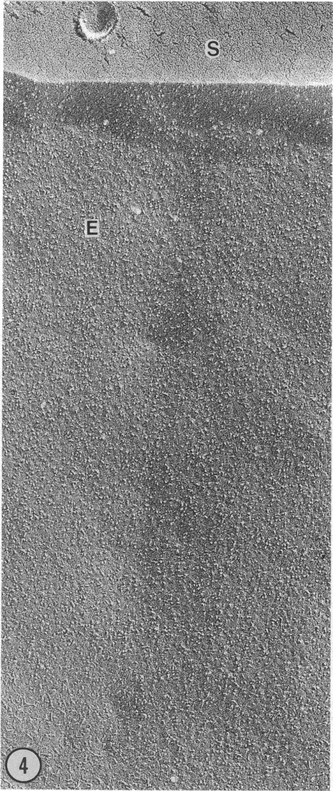
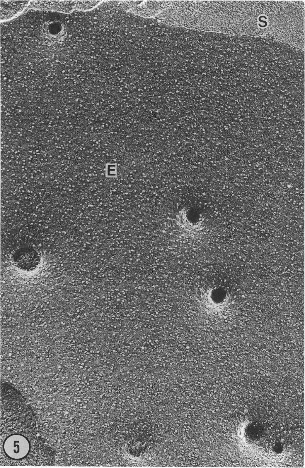
Left (labeled #4) - external surface of an untreated red blood cell. Magnification is 60,000X. Right (labeled #5) - External surface of an electropermeabilized human red blood cell 40 ms after the application of the pulses. Magnification is 60,000X. Figures and captions were modified from Chang and Reese, 1990. Biophys J. Vol 58, p 01-12.
But how does this relate to a treatment for atrial fibrillation? Well it turns out the goal of Pulsed Field Ablation for arrhythmia is to cause such extreme electroporation of the aberrant cardiac cells that they just die. Without the badly behaving cells, the heart can then rely on its healthy cells to keep the beat.
It’s not difficult to imagine how electroporation could kill a cell after seeing the images above. If enough of those pits form there would barely be any membrane left to hold the cell together! Or perhaps the pores allow molecules into the cell that shouldn’t be there, ultimately leading to the cell’s demise. Or vice versa, perhaps critically important cell components exit the cell through the pores and the cell can’t recover from the loss. Another theory is that the flux of charged particles through the pores, like potassium or sodium, actually generates resistive heat and the increased temperatures cause enough damage to kill the hole-punched cells. All of the above may happen to the cardiac cells that are targeted during a PFA treatment.
One of the purported benefits of PFA to other ablation techniques is that it leaves nerve and blood vessel tissues near the stimulated site intact. It’s not entirely clear why, but my hypothesis is that different cell types have membranes that are so chemically variable that they just respond differently to the same electrical fields. Cells in different tissue types have wildly different cell membrane compositions; some have higher fractions of cholesterol, some are thickly coated in sugars, and they each contain a different set of proteins. Many of the molecules that make up the cell membrane will reorient in space when exposed to an external electric field and the way they do will depend on their exact structure, charge, environment, etc. Oh and all of these features are dynamic in time too, so understanding why some cells behave one way in an electrical field and other cells behave another way is not trivial. There are several completed and upcoming clinical trials testing electroporation as a treatment for cancer. It would be interesting to learn how different the pulse protocols need to be when targeting a tumor vs. a myocardium. I'm sure an army of scientists and clinicians are all over it!
Further reading:
If you want to explore why pores form in the membrane where they do, check out this review of some of the theories.
If you want to read more about the FARAPULSE clinical trial, check out the clinical trial data or read Boston Scientific’s press release.
Comments
Post a Comment